

What Can Digital Implant Planning Change That a Traditional Implant Plan Cannot?
Quick Summary
- Restoration-first approach: Digital planning designs your ideal final tooth before the surgical implant is placed, ensuring proper aesthetics and bite alignment.
- Overcoming 2D limits: Traditional two-dimensional X-rays cannot reveal bone width or precise nerve pathways; three-dimensional scans eliminate that guesswork.
- Anatomical protection: Virtual simulation maps bone density and vital structures in advance, making the physical surgery calm, targeted, and controlled.
- Informed decision-making: At the practices of Dr. Alex Fneiche in Brussels and Trazegnies, digital assessment clarifies timelines, candidacy, and transparent costs before treatment begins.
Replacing a missing tooth is more than securing a small titanium post into the jaw. The genuine objective is recreating a natural tooth that balances your bite, protects your oral biology, and functions comfortably for decades.
Digital implant planning fundamentally changes this process. It replaces manual estimation with computer-guided architectural precision, ensuring every clinical decision is tested and refined before treatment begins.
Digital Planning Is Not Just About Using More Technology
Advanced equipment offers little benefit if it merely serves as a high-tech display. In modern dentistry, digital planning is not about adding unnecessary complexity.
It represents a deliberate clinical philosophy. The focus shifts from reacting to anatomical challenges during surgery to resolving them systematically in advance.
The Difference Is How the Implant Is Planned
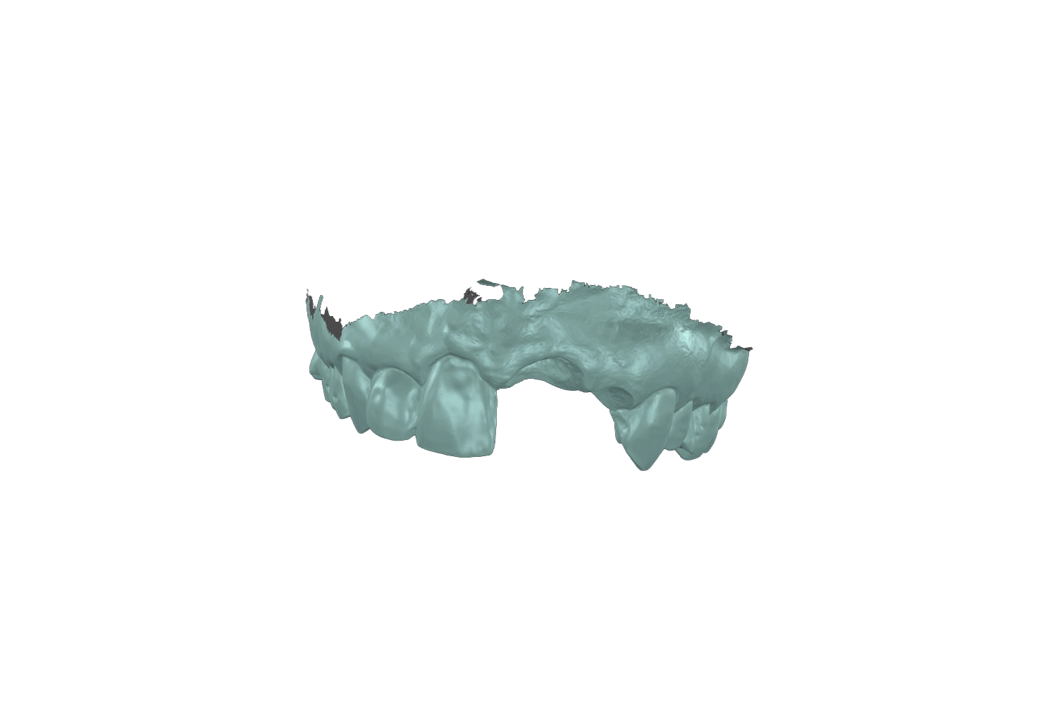
In conventional implant treatment, crucial spatial decisions often take place during the surgical appointment. The clinician reflects the gum tissue and assesses the exposed bone directly in the operatory chair.
Digital planning moves that critical evaluation to the diagnostic stage. By analyzing your bone volume and dental relationships on a virtual model beforehand, potential obstacles are identified and resolved before surgery. This pre-procedural clarity creates a calm, structured, and efficient appointment.
The Final Tooth Can Be Considered Before the Implant Is Placed
Historically, dental implants were placed wherever bone volume was easiest to find. The artificial crown was then shaped later to accommodate wherever that post emerged.
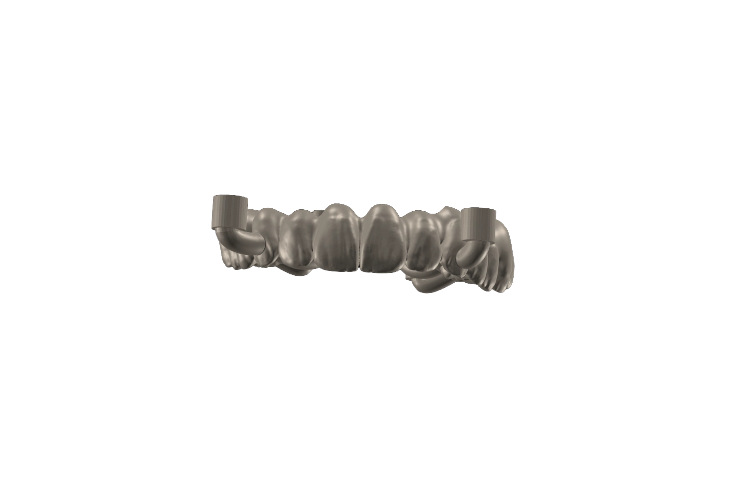
Digital workflows reverse that order entirely through prosthetically driven backwards planning. Dr. Alex Fneiche determines the ideal shape, contour, and biting position of your future tooth first. The implant is then positioned specifically to support that definitive restoration.
What Can Digital Implant Planning Actually Change?
Moving from static assumptions to three-dimensional modeling directly influences how your surgery is organized and executed.
By analyzing the full oral environment, digital planning actively alters three critical clinical elements.
It Can Change Where the Implant Is Placed
An implant must reside entirely within healthy bone while maintaining a safe distance from neighboring tooth roots.
Digital simulation allows a clinician to evaluate multiple placement angles down to fractions of a millimeter. This ensures the fixture is placed at the exact depth and angle required to distribute everyday chewing forces evenly.
It Can Reveal Anatomical Limitations Before Surgery
The human jaw houses delicate anatomical landmarks, including the inferior alveolar nerve in the lower jaw and the maxillary sinuses in the upper jaw.
Standard clinical exams cannot confirm the exact spatial depth of these boundaries. Three-dimensional planning makes these structures clearly visible in cross-section, substantially reducing procedural uncertainty.
It Can Change Whether Additional Treatment Is Needed
Patients are occasionally told they lack adequate bone based solely on a flat, two-dimensional radiograph.
Comprehensive 3D scans frequently reveal that bone thickness is adequate when viewed from a different angle. In other cases, digital planning identifies precisely where targeted bone or gum grafting is necessary, eliminating unexpected surprises during the procedure.
Why Does Implant Position Matter So Much?
A dental implant serves as the foundation for your new tooth. Much like building architecture, the alignment of the base dictates the performance of the visible structure.
Even slight variances in placement can create long-term biological and functional consequences.
The Implant Position Influences the Final Restoration
The angle and depth of an implant dictate how the custom crown connects to the foundation.
If an implant is placed at an awkward angle, the crown must be artificially contoured to compensate. This can create unnatural contours that trap food particles and complicate daily cleaning. Proper alignment allows your final crown to emerge seamlessly from the gum line.
Small Differences in Position Can Have Larger Consequences
A placement discrepancy of one or two millimeters can affect how biological tissues maintain themselves over time.
- Implants placed too close to natural roots can compromise adjacent teeth.
- Implants placed with insufficient surrounding bone risk future gum recession.
- Implants positioned with sufficient biological margins promote stable osseointegration and protect long-term tissue health.
The Goal Is Not to Place the Implant Where the Bone Happens to Be
The primary function of a tooth replacement is restoring comfortable chewing. Placing an implant simply where bone is thickest can compromise how the upper and lower teeth meet.
The genuine objective is placing the implant where it delivers optimal mechanical stability and balance. If natural bone is deficient in that exact position, the site is prepared properly rather than compromising the restoration.
What Traditional Implant Planning Can and Cannot Tell You
Traditional diagnostic tools retain clinical utility, but recognizing their limitations helps explain why digital planning has become an indispensable standard.
Traditional Planning Still Has a Place in Implant Treatment
Conventional methods, including physical impressions, dental study models, and two-dimensional radiographs, remain valuable introductory tools.
They provide an accessible baseline assessment of your periodontal health, active decay, and overall bite alignment. They help your dentist determine if dental implant therapy is a realistic option for you.
Two-Dimensional Information Has Limits
Standard panoramic X-rays compress a curved, three-dimensional jaw into a single flat image.
A flat X-ray shows bone height, but it cannot show bone width, ridge density, or cross-sectional contours. Relying strictly on two-dimensional images means critical anatomical details remain hidden until the surgical phase begins.
Digital Planning Adds Another Layer of Information
Three-dimensional scans capture the complete volume of your jawbone, soft tissue, and dental arches simultaneously.
A clinician can rotate your jaw anatomy along any plane and evaluate virtual placement before touching a surgical instrument. This replaces clinical estimation with verifiable anatomical information.
How Dr. Alex Fneiche Uses Digital Implant Planning
In his practices in Brussels and Trazegnies, Dr. Alex Fneiche organizes implant treatment around clinical predictability and patient clarity.
Planning Starts With the Final Restoration
Every treatment plan begins with your final smile in mind. Dr. Fneiche establishes the dimensions, natural proportions, and functional contact points of your future crown first.
Designing the visible restoration at the start ensures that every diagnostic and surgical step serves a deliberate functional goal.
Bone, Soft Tissue and Anatomy Are Assessed in Three Dimensions
Using comprehensive 3D imaging, Dr. Fneiche evaluates your anatomy in high resolution.
This diagnostic phase examines bone volume, root spacing, and tissue density relative to your occlusal dynamics. By analyzing your anatomy thoroughly, he confirms whether your case is straightforward or requires supportive preparatory care.
The Implant Position Is Planned Around the Whole Treatment
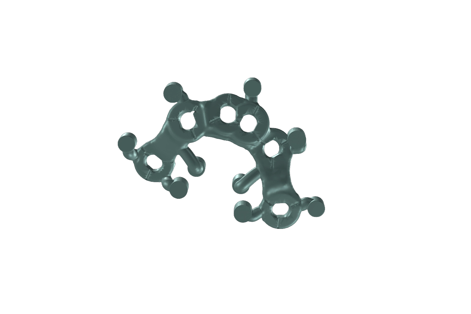
When appropriate, Dr. Fneiche utilizes custom surgical guides to transfer the digital blueprint directly into clinical practice.
This structured workflow supports minimally invasive techniques, controlled fixture placement, and predictable tissue healing. Detailed clinical documentation is maintained throughout your file, ensuring transparent tracking from your first consultation to your final restoration.
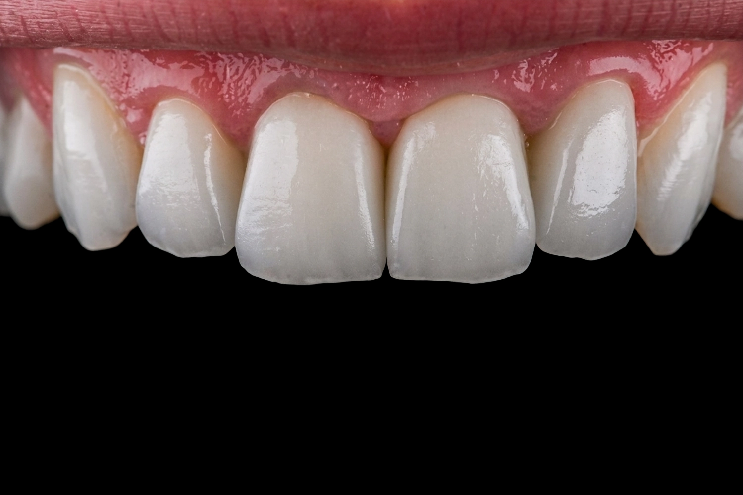
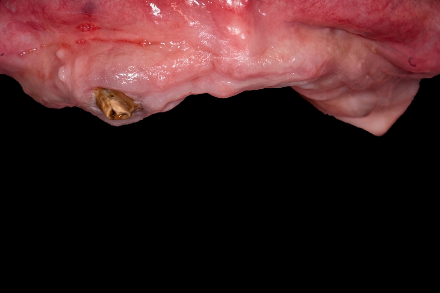
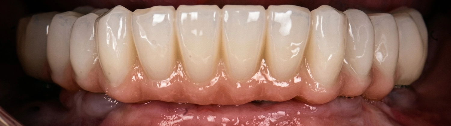
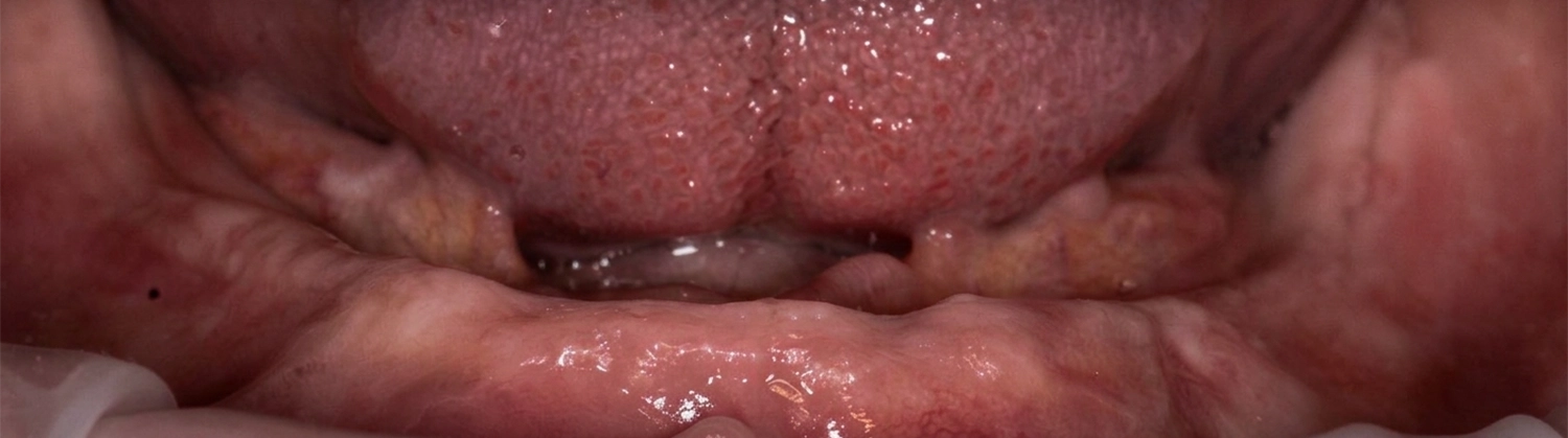
What Does This Mean for You?
Choosing a digitally planned approach changes how your treatment feels from start to finish.
You Are Not Simply Planning Where the Implant Goes
You are not merely receiving a standalone dental procedure.
You are entering a structured treatment pathway where every stage is planned in advance. You receive an upfront written plan, realistic timelines, and clear fee guidance, with single implant pricing starting from 900 euros and exact costs confirmed after your full assessment.
You Are Planning the Tooth, the Biology and the Long-Term Result
A digitally planned implant is designed to protect your quality of life for the long term.
By respecting natural biological boundaries and balancing functional forces, this approach supports smooth osseointegration and natural aesthetics. The final result is a tooth that looks natural, feels stable, and restores your confidence when eating and speaking.
What Should You Do If You Are Considering Dental Implant Treatment?
Understanding your treatment options begins with gathering accurate information about your unique dental anatomy.
Start With an Assessment of Your Individual Anatomy
Because every smile presents distinct biological conditions, generalized answers can only take you so far.
A comprehensive consultation with Dr. Alex Fneiche in Brussels or Trazegnies provides complete diagnostic clarity. Together, you will review your 3D assessment, discuss whether a same-day temporary tooth is possible for your situation, and explore a structured treatment plan tailored to your needs.
Schedule your consultation today to explore your options with clarity, precision, and complete confidence.
Schedule your consultation todayFrequently Asked Questions (FAQ)
1. What makes 3D digital implant planning safer than traditional methods?
Traditional implant planning relies largely on two-dimensional X-rays, which only display bone height while leaving bone width and nerve depth hidden. 3D digital planning uses high-resolution diagnostic imaging to reconstruct the full volume of your jawbone, adjacent tooth roots, and vital structures, such as the inferior alveolar nerve and sinus cavities. By evaluating these biological boundaries prior to the appointment, your clinician can precisely determine the placement angle and depth, significantly minimizing surgical surprises and protecting surrounding tissues.
2. Why does Dr. Alex Fneiche plan the crown design before placing the implant?
This approach is known as prosthetically driven backwards planning. Rather than inserting an implant wherever bone is easiest to access and forcing an artificial crown to fit on top, the process begins by designing your ideal visible tooth. The implant fixture is then positioned specifically to support that definitive restoration. This ensures optimal chewing alignment, prevents unnatural contours that trap food, and delivers a natural aesthetic emergence from the gumline.
3. How do surgical guides improve recovery and procedure comfort?
A 3D surgical guide acts as a custom blueprint created from your digital treatment plan. It fits securely over your teeth or gums during surgery, directing the instruments at the exact depth and trajectory planned in the software. Because key decisions have already been resolved digitally, the surgical procedure is streamlined, targeted, and often minimally invasive. This controlled technique helps reduce unnecessary tissue disruption, which supports more comfortable healing and smoother recovery.
4. Can I get a dental implant if I have been told I have low bone density?
A flat, two-dimensional X-ray can sometimes underestimate bone thickness due to projection angles. A comprehensive three-dimensional assessment evaluates the entire cross-section of your ridge and may reveal adequate bone volume that was not visible on standard images. If bone or gum tissue is genuinely insufficient, digital planning allows Dr. Fneiche to identify the exact location and volume required for targeted bone or soft tissue grafting before or during the procedure.
5. Is it possible to receive a temporary tooth on the same day as surgery?
In selected clinical cases, a temporary tooth can be fitted on the same day the implant fixture is placed. Candidacy for immediate provisional restoration depends on several diagnostic criteria, including your primary bone stability, bite dynamics, and tissue health. During your initial 3D digital assessment, Dr. Alex Fneiche will determine whether same-day loading is clinically safe and suitable for your specific anatomy.
6. How much does a dental implant cost, and what does the fee cover?
At our practices, treatment starts from €900 for a single implant fixture. Because every patient presents a unique anatomical profile, the total cost depends on factors such as diagnostic 3D imaging, surgical guides, preparatory bone or tissue grafting (if necessary), and your final custom crown. Following your comprehensive digital consultation, you will receive a transparent, itemized written treatment plan outlining every milestone and fee with no hidden costs.
7. Where are your practices located, and do you accept patients from neighboring areas?
Dr. Alex Fneiche welcomes patients across two clinical locations: one in Brussels and one in Trazegnies (Courcelles).
- Our Brussels practice regularly welcomes patients from surrounding municipalities and neighborhoods such as Ixelles, Etterbeek, Uccle, Woluwe-Saint-Lambert, Schaerbeek, and the wider Brussels-Capital Region.
- Our Trazegnies clinic frequently provides care for patients traveling from nearby towns and districts across Hainaut, including Charleroi, Courcelles, Chapelle-lez-Herlaimont, Manage, Gosselies, and Fontaine-l’Évêque.
Both locations are equipped for complete digital diagnostic assessments and advanced restorative care.
8. How long does the complete digital implant journey take from start to finish?
While the surgical placement itself is efficient and completed in a single appointment, true biological osseointegration (the natural bonding between the biocompatible implant post and your jawbone) typically takes between 8 and 16 weeks, depending on individual healing and whether grafting was performed. Once integration is confirmed through clinical monitoring, your final custom crown is placed to complete your permanent, fully functional smile.